
Radiography Imaging: Medical imaging technologies are an important aspect of helping healthcare professionals to diagnose and treat illness. Radiography is a method of imaging that enables healthcare professionals to see the internal structure of the human body.
Table of Contents
Radiography is perhaps the most frequently used imaging modality available to create diagnostic images of bones, soft tissues and organs in order for healthcare professionals to quickly and accurately diagnose patients who may have suffered from burns, fractures, infections or disease.
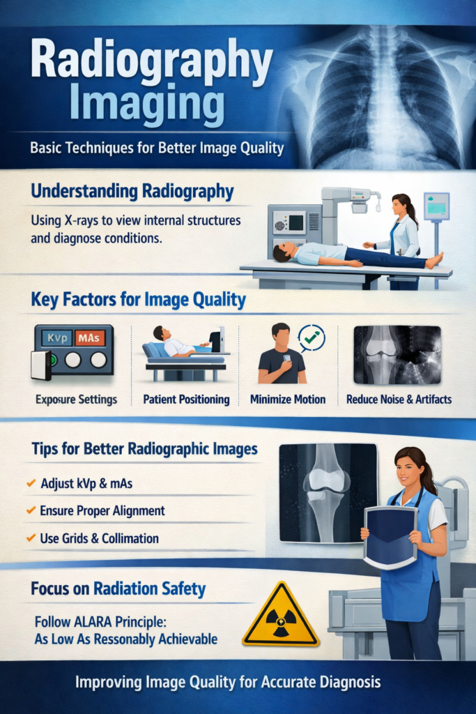
Because radiographs are used so frequently in the diagnosis of patients, radiographers have to fully understand how much image quality matters when it comes to making an accurate diagnosis for patients. Quality images assist radiologists in determining whether or not a patient has an illness and provide them with the tools needed to develop an appropriate treatment plan in a timely manner. Conversely, poor quality images may lead to inaccurate diagnoses or unnecessary repeat studies, which is why it is critical that medical imaging professionals understand the fundamental principles of how to improve the quality of Radiography Imaging.Similarly, radiological image quality depends on characteristics of imaging equipment, skill of the radiological technician, radiation exposure to the patient, and imaging time. Five major components of medical imaging process are –
The object in the patient body
The imaging equipment
Radiographer
Image itself
The observers or radiologist who will conclude the study
Understanding Radiography in Modern Medicine
So you want to know about the techniques that improve image quality in radiography. Well first you need to understand what radiography is about. Radiography Imaging uses X-rays to take pictures of what’s inside the human body. When these X-rays go through the body they get absorbed by tissues at different rates.
For instance bones absorb a lot of radiation which’s why they look white in the pictures. Soft tissues look gray. Areas with air like the lungs look darker. This difference in how tissues absorb radiation helps doctors see what is going on inside the body.
Radiography is used in lots of places like hospitals, diagnostic centers and clinics. Doctors use it to find out if someone has a broken bone, a lung infection, a dental problem or many other health issues. Radiography is really helpful for figuring out what is wrong, with someones body
Quality Imaging
Quality imaging is a part of medical imaging. It is about how the pictures show the bodys structures. In words it is about how much the picture looks like the real thing. To get quality imaging you need to use the equipment correctly. You also need to choose the techniques. When you do these things right the pictures are clearer and more helpful for doctors.
Impact of Quality Defects
When the quality of the pictures is not good it can cause a lot of problems. If the pictures are not clear it takes doctors longer to understand what is going on. Sometimes they need to take the pictures which means the patient gets more radiation. This also means work for the medical staff. If the pictures are not clear it can be hard for doctors to make the decisions, about treatment. Quality imaging is important because repeat pictures cost money and can delay the patient getting the right treatment. Quality imaging is what doctors need to make sure patients get the care.
What Makes a Good Radiographic Image
When you look at a medical image, you want it sharp and useful, right? A handful of things shape how good that image actually turns out. Let’s talk about the big six.
Also Read: How to Maintain a Healthy Lifestyle During Exam Preparation
Spatial Resolution
Spatial resolution is basically how well the system can tell apart two tiny objects sitting close together. If the spatial resolution is high, you’ll see crisp, detailed images, making it easy to spot small anatomical features. Think about trying to see exactly where bone stops and soft tissue begins — good spatial resolution makes that boundary obvious.
Contrast Resolution
Then there’s contrast resolution. This one’s about picking out differences between tissues that look almost the same, at least density-wise. Take the liver and spleen, or fat and muscle — they don’t always stand out from each other. But if the system’s contrast resolution is up to snuff, you can spot even minor differences, so radiologists can catch abnormalities early on.
Noise
Noise is that annoying graininess you see when an image just doesn’t look smooth. It pops up as random patterns that mess with the clarity, making it tough to pick out fine details. Too much noise? Good luck spotting anything subtle. That’s why cutting down on noise really boosts both the contrast and the overall quality of the image.
Artifacts
Artifacts are fake features—stuff on an image that isn’t actually part of the body. They usually show up because of equipment glitches, the patient moving, or little mistakes during imaging. The problem is, artifacts can hide the things you actually need to see, which makes diagnosing harder. So, getting rid of artifacts matters a lot if you want reliable, accurate radiographs.
Distortion
Distortion is what happens when body parts look the wrong size, shape, or in the wrong place on an image. The goal in medical imaging is to keep everything true to life—real dimensions, real positions. Distortion sneaks in when the patient isn’t positioned right, the machine isn’t lined up, or the equipment itself has limits.
Compromises in Image Quality
In medical imaging, you almost never get perfection. Boosting one part of image quality often means sacrificing another. Say you sharpen the image—you might end up with more noise. Or if you change the exposure to get better contrast, you might lose some detail. That’s why radiologic technologists are always making trade-offs, adjusting settings to find the best mix of visibility, sharpness, and safety for the patient.
How to Get Better Radiographic Images
If you want sharp, useful radiographic images, you need more than just book knowledge. It’s part science, part hands-on skill. Here’s what works in real practice.
Start with exposure—get it right for the patient and the body part you’re looking at. Don’t just use the same settings for everyone. Change them up so the image actually shows what you need, with good contrast and brightness.
Next, pay attention to how everything lines up. The X-ray tube, the patient, and the detector all need to be in the right spot. If they’re not, you’ll end up with weird distortions or patches that aren’t clear.
And don’t forget the gear. Equipment that’s well cared for runs better and gives you images you can trust. Make a habit of checking and maintaining it, not just when something breaks.
Digital imaging has made things easier, too. Now you can tweak images on a computer—fix brightness, bump up contrast, sharpen edges. But don’t overdo it. Too much editing can actually hide important details, and that’s the last thing you want.link
Why Equipment Maintenance Matters
If your equipment’s not working right, your images won’t be either. Worse, bad equipment can mean patients get more radiation than they need.
Stay on top of maintenance. Check X-ray tubes and detectors, update software, and run quality control tests. When everything’s in top shape, you get clear images and safer exams every time.
Staying Safe Around Radiation
Radiography is powerful, but it comes with risks. Ionizing radiation isn’t something to mess around with, so safety always comes first.
That’s why everyone in the field follows the ALARA principle—keep radiation “As Low As Reasonably Achievable.” Use shields, set exposures carefully, and work efficiently to protect both patients and staff.
How Technology Keeps Changing Radiography
Radiographic imaging has come a long way. Film is mostly history. Digital systems are fast, let you adjust images on the fly, and make sharing results with other doctors a breeze.
AI is starting to help, too. It can scan images for signs of fractures or lung problems, making it easier for doctors to spot issues quickly and accurately.
Radiography just keeps getting better. As the technology grows, expect scans to get even clearer, quicker, and more widely available.
Conclusion
Radiographic imaging has become an important tool in modern medicine because it allows medical professionals to identify many different ailments quickly. Obtaining quality images requires skill and patient cooperation. Each examination has its own set of steps in terms of setup and patient positioning; each scenario requires its own considerations.
Thanks to the efforts of the medical professionals and patient understanding of the importance of safety and proper imaging technique; valuable images are generated for diagnostic quality.
Not only will technologists help you, but medical imaging technology continues to improve with advancements in each of the three imaging modalities. The most important advancement is the reduction of radiation exposure to the patient.